
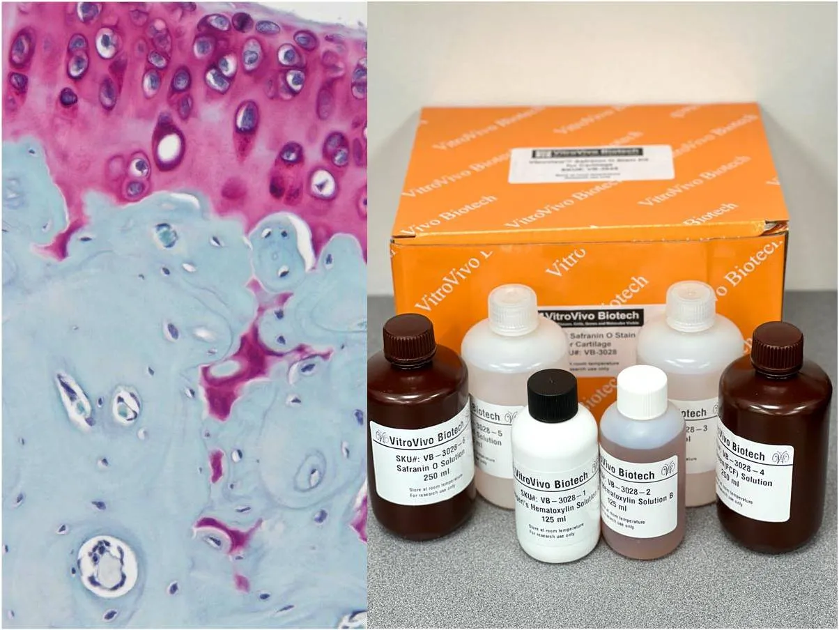
Osteochondral research often asks two questions at once: how well cartilage matrix is preserved and how actively the underlying bone is being remodeled. safranin o staining helps researchers visualize proteoglycan-rich cartilage, providing a practical way to compare matrix retention across experimental groups.
That structural information becomes more useful when it is paired with a cellular measure from the adjacent bone compartment. Instead of treating cartilage and subchondral bone as separate systems, researchers can examine how changes on one side relate to activity on the other.
Map Matrix Preservation First
Cartilage contains abundant proteoglycans that contribute to hydration and resistance to compression. Safranin O creates strong contrast in proteoglycan-rich regions, allowing investigators to compare intact matrix with areas showing reduced staining.
To study the bone side of the same interface, a TRAP stain kit osteoclast can be used to identify tartrate-resistant acid phosphatase activity. Multinucleated TRAP-positive cells are commonly evaluated as osteoclasts in bone-remodeling research.
Keep the Endpoints Distinct
Proteoglycan loss and osteoclast activity describe different biological events. Reduced cartilage staining does not automatically prove increased bone resorption, and more TRAP-positive cells do not by themselves establish that cartilage matrix has deteriorated.
The strongest study design measures each endpoint independently before comparing patterns. Researchers can then ask whether matrix depletion and osteoclast activity occur in the same region, at different time points, or in response to different experimental conditions.
Plan Section Orientation Carefully
The bone–cartilage interface is highly dependent on orientation. Sections cut at inconsistent angles can change the apparent cartilage thickness, growth-plate appearance, or amount of subchondral bone visible in a field.
Specimens should therefore be embedded and sectioned using repeatable anatomical landmarks. A consistent cutting plane makes it easier to compare corresponding regions between treatment groups and reduces variation caused by sampling rather than biology.
Consider Decalcification Early
Bone-containing specimens often require decalcification before routine paraffin sectioning. The chosen method should preserve enough morphology and chemical activity for the stains planned later in the workflow.
Researchers should document decalcification conditions carefully because prolonged or harsh treatment can affect tissue components. When enzyme histochemistry is important, processing should be planned with preservation of the relevant activity in mind.
Use Serial Sections Strategically
Adjacent sections are useful when two stains require different chemistries. One level can be reserved for cartilage matrix assessment, while a neighboring level is used for osteoclast-focused histochemistry.
A section map can record slide number, cutting depth, orientation, and intended assay. This simple step helps investigators compare related regions later and prevents limited osteochondral tissue from being used inefficiently.
Define Regions Before Scoring
Quantification becomes more reliable when anatomical regions are chosen before the full dataset is reviewed. Researchers may divide cartilage into superficial, middle, and deep zones or define specific areas of subchondral bone.
For osteoclast analysis, counting rules should state which cells qualify, where they are counted, and whether results are expressed per area, bone surface, or another predefined reference. Consistent rules reduce observer-driven variation.
Standardize Imaging Conditions
Microscope settings can change how staining intensity and cell morphology appear. Magnification, illumination, exposure, white balance, and digital processing should remain consistent when images will be compared across experimental groups.
The same principle applies to image-analysis software. Thresholds, regions of interest, exclusion rules, and measurement settings should be established before final analysis so technical choices do not shift from one sample to another.
Add Routine Morphology for Context
Special stains are most informative when researchers can relate them to overall tissue architecture. An adjacent H&E section can help identify fractures, tissue folds, inflammatory areas, vascular changes, or other features that may affect interpretation.
This broader context can explain why two regions with similar special-stain intensity behave differently. It also provides a useful reference when matching cartilage and bone features across serial sections.
Conclusion
Osteochondral remodeling cannot be understood from one tissue component alone. Cartilage matrix preservation and osteoclast activity represent separate but potentially related processes that benefit from complementary histological assessment.
By controlling orientation, decalcification, section allocation, scoring, and imaging, researchers can compare these endpoints more confidently. A coordinated workflow turns neighboring sections into a connected view of how cartilage and underlying bone respond within the same experimental model.